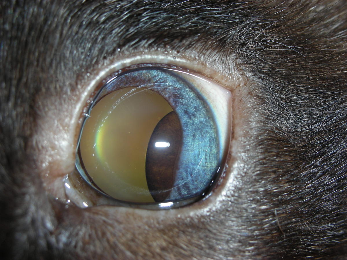
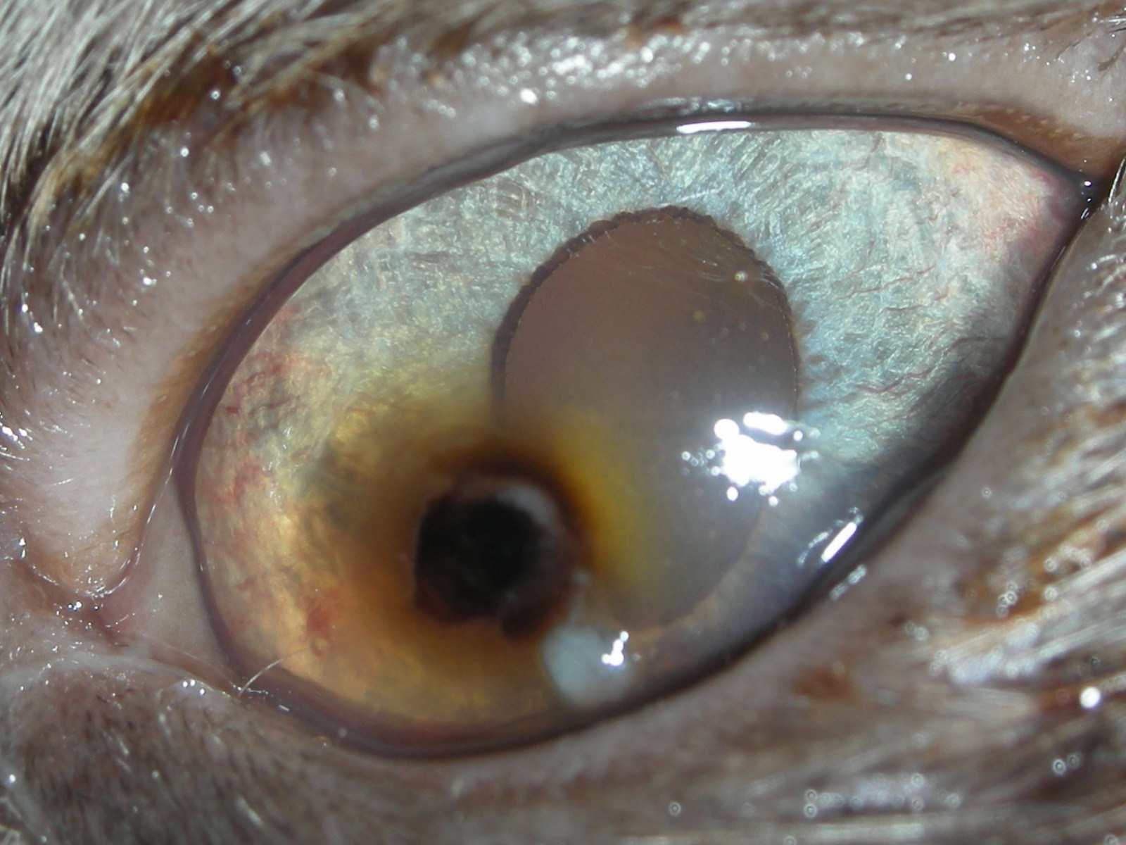
Benign uveal cyst in the posterior chamber of a cat's eye

Benign uveal cyst in the anterior chamber of a dog's eye
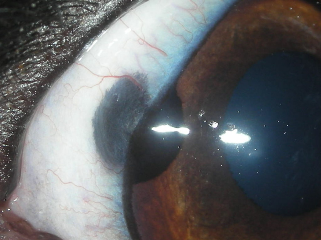
Benign growth although therapy is required to save the eye including surgical resection, cryotherapy, diode or carbon dioxide laser therapy.

Diode laser therapy for solitary, defined lesions can often be curative.
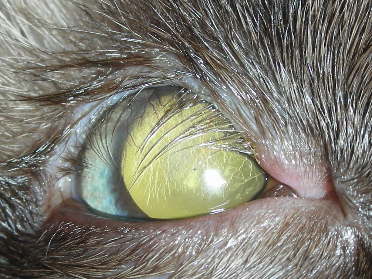
Cryoepilation (permanent hair removal) along the missing eyelid margin often relieves the corneal trauma and pain associated with this condition.
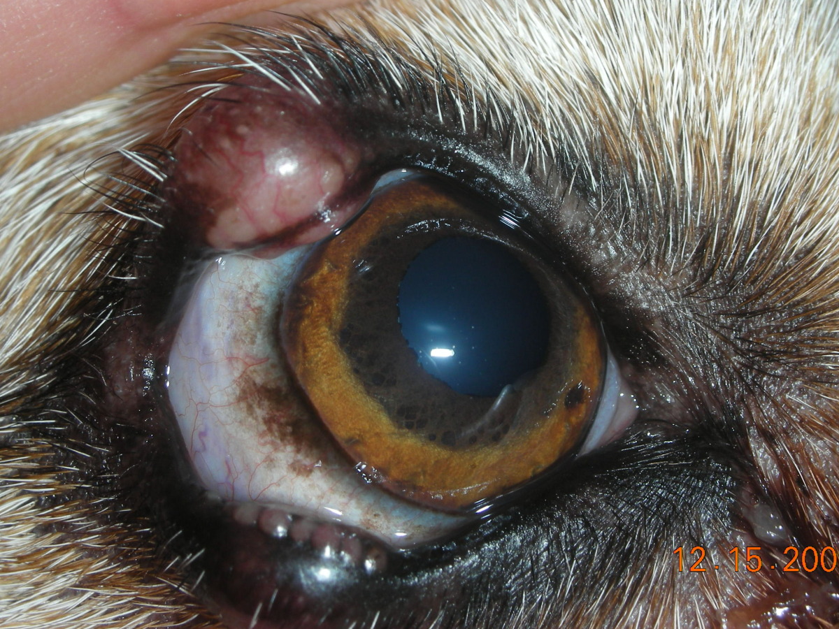
A treatable immune-mediated disease that often requires long-term or life-long therapy.
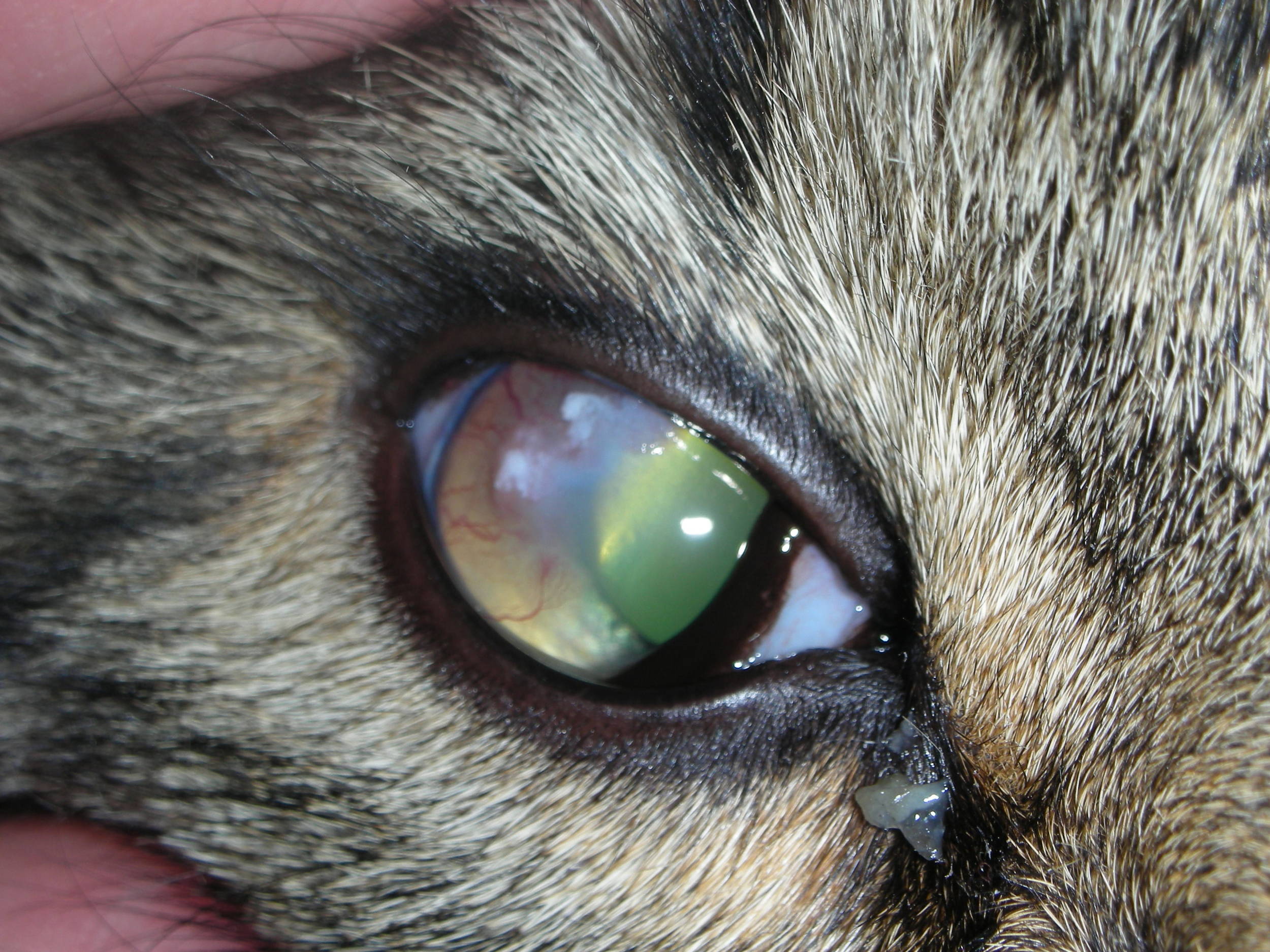
Not a painful disease although patients require therapy to prevent vision loss.

Usually a benign age related ocular degeneration.
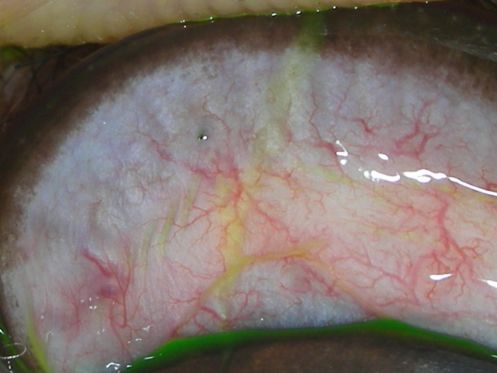
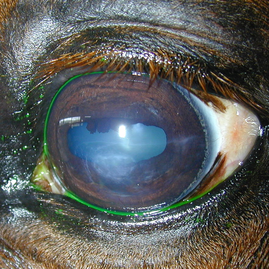
Very painful and can lead to loss of the eye if a deep corneal ulceration develops.
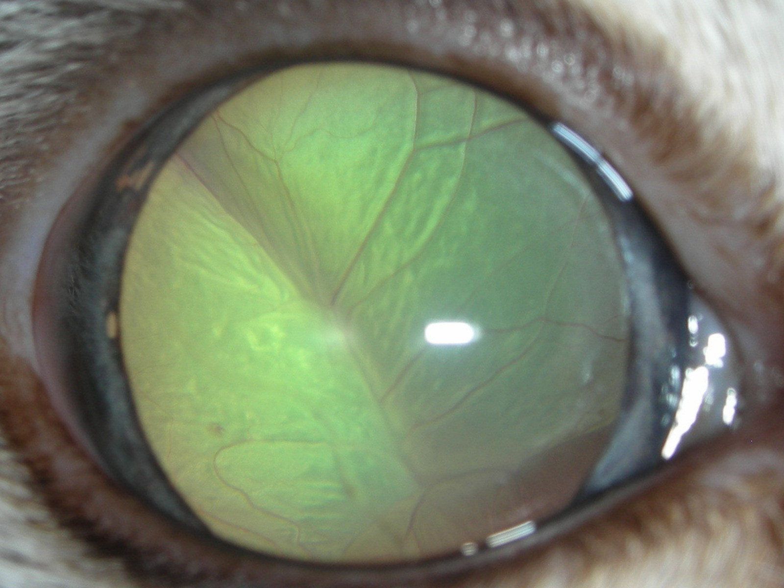
Typical appearance of a complete serous retinal detachment. If appropriate and rapid therapy is instituted patients often have a favorable visual outcome.

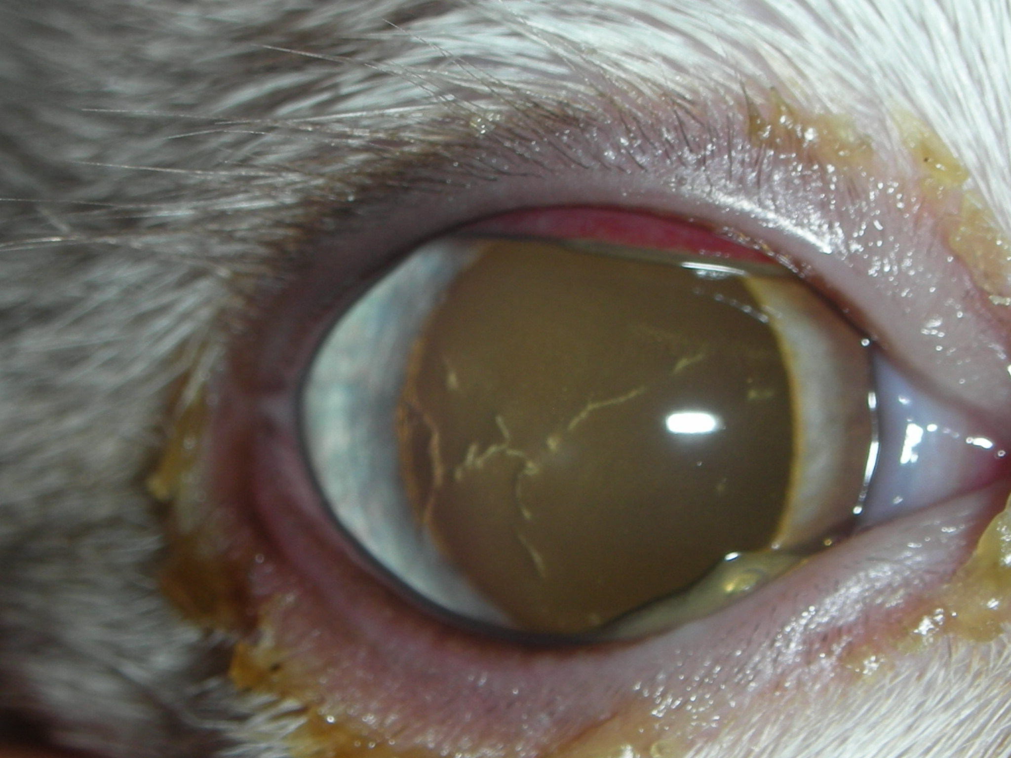
One of the most common causes of feline corneal ulceration.
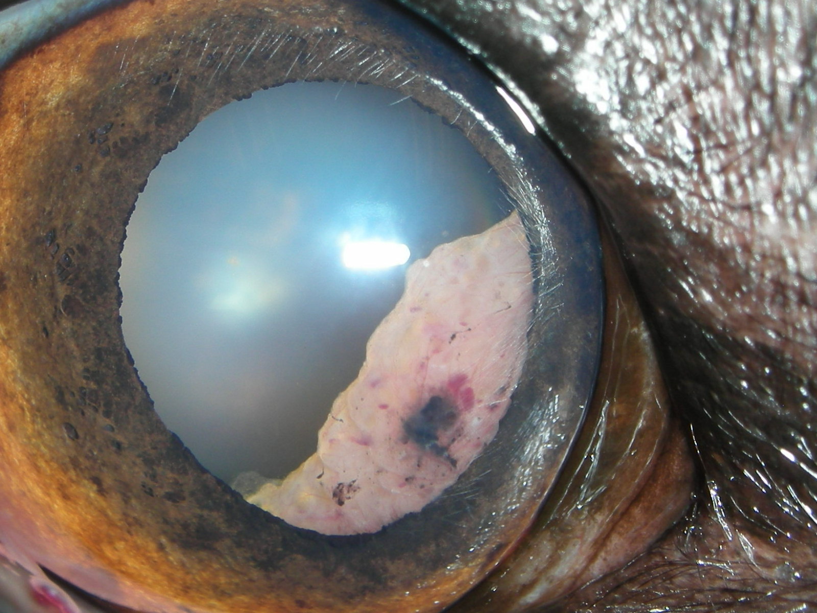
Surgical removal (superficial keratectomy) +/- combined with a corneal graft is often required.
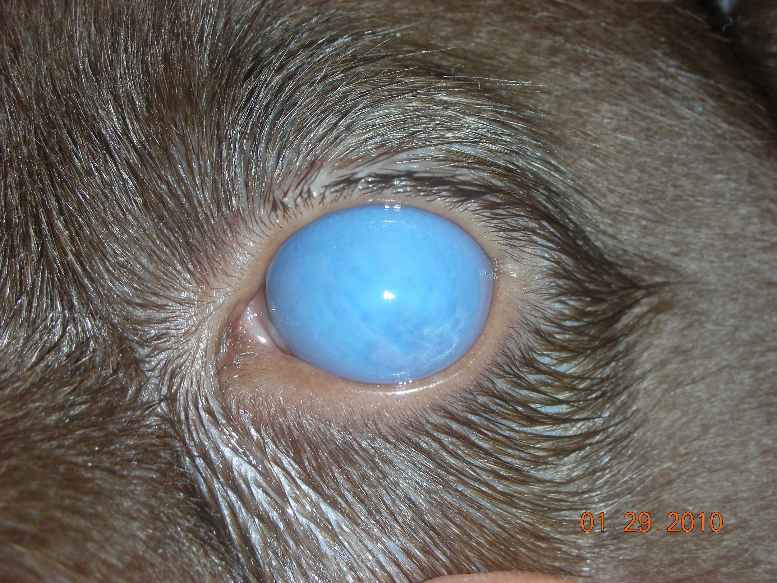
Fortunately this occurs very rarely with modern vaccines. Unfortunately this condition can lead to secondary glaucoma and loss of the eye.
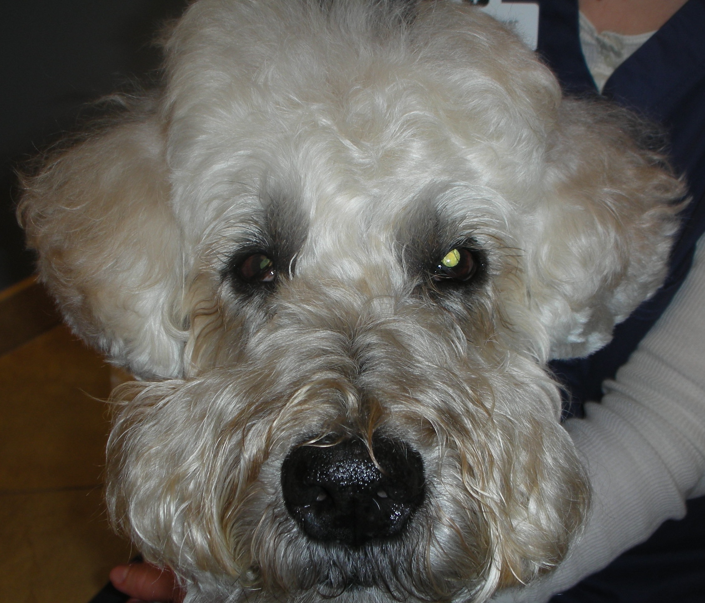
Most often a benign and spontaneously resolving neurological disorder.
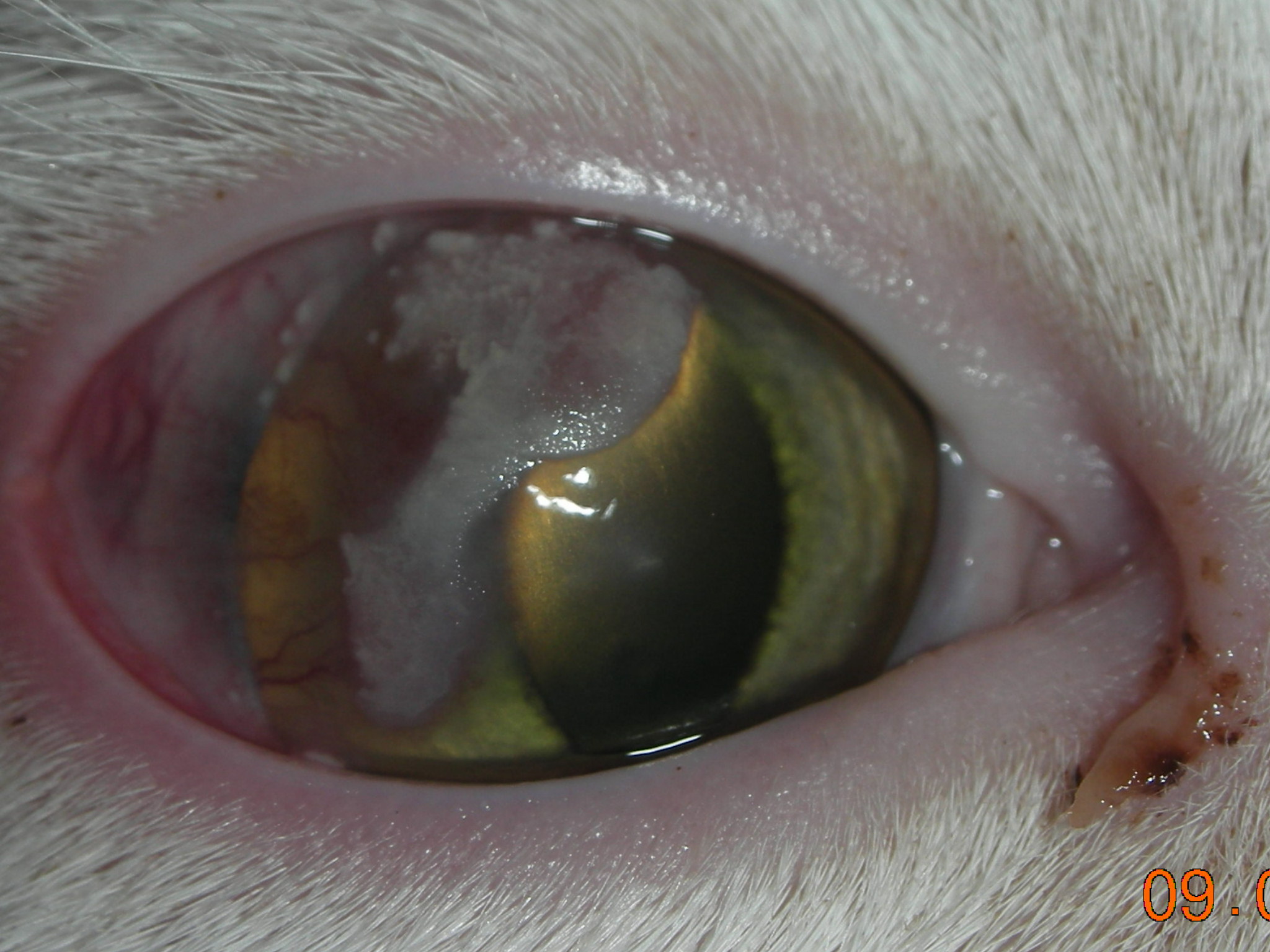
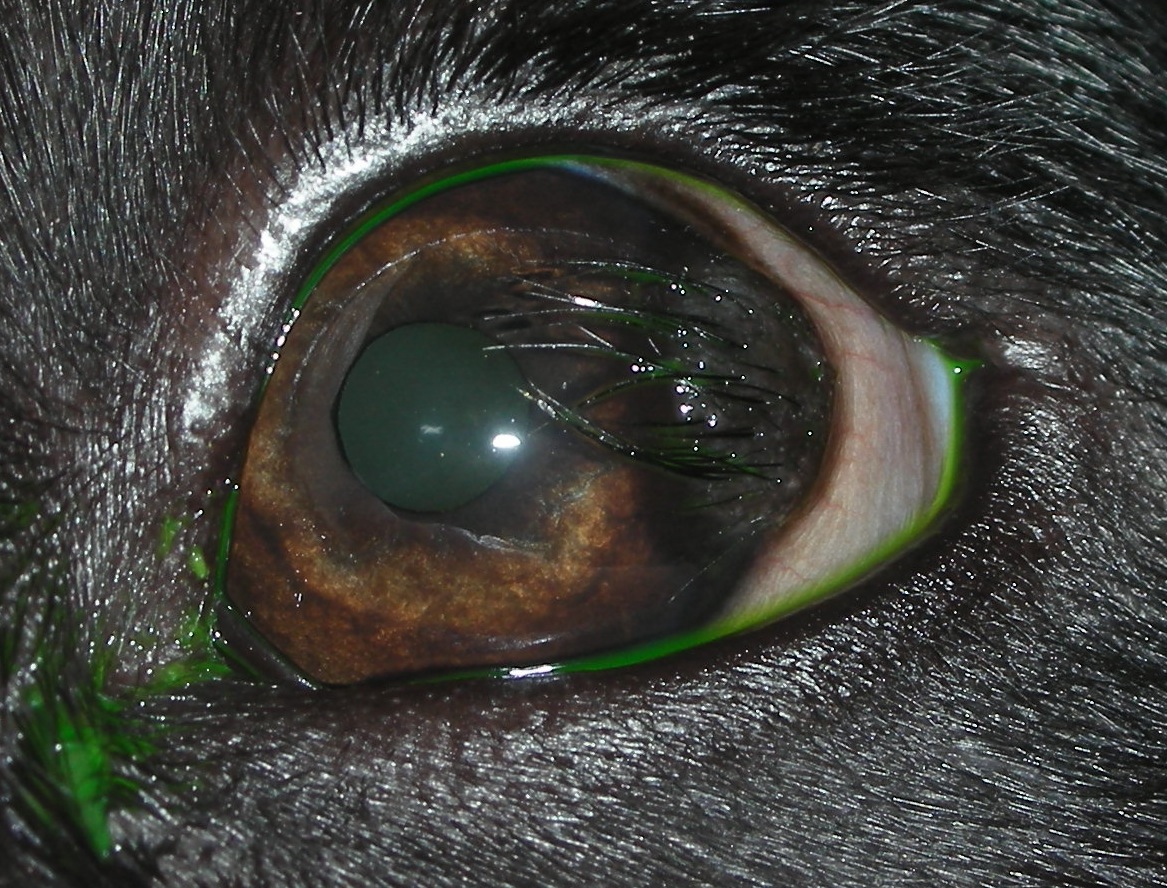
A congenital abnormality: normal tissue (containing hair follicles) growing in an abnormal location (eg. the cornea, conjunctiva or eyelid). Surgical removal +/- grafting surgery is often indicated to relieve patients of the discomfort associated with the hairs rubbing on the surface of the eye.
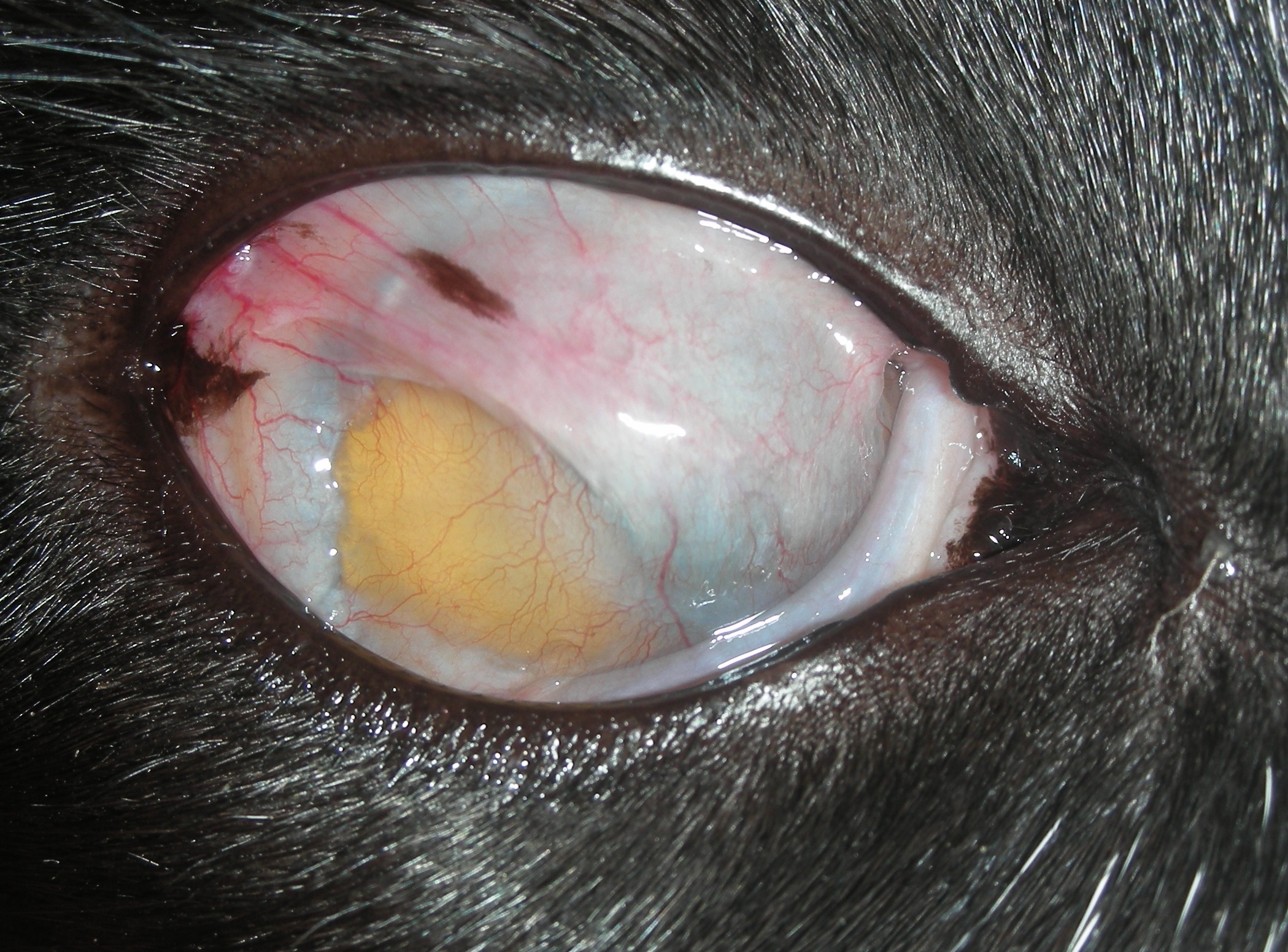
Fusion of conjunctival tissue to the surface of the cornea. Indicates early life complication of a feline herpes virus infection.
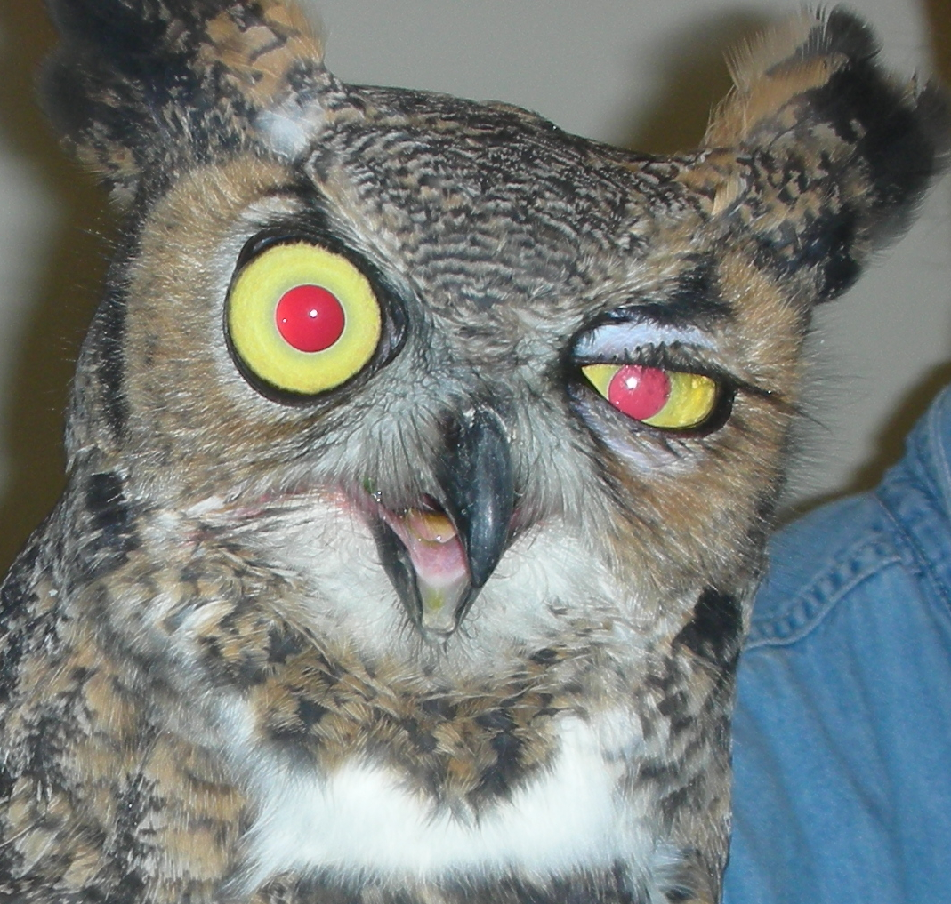
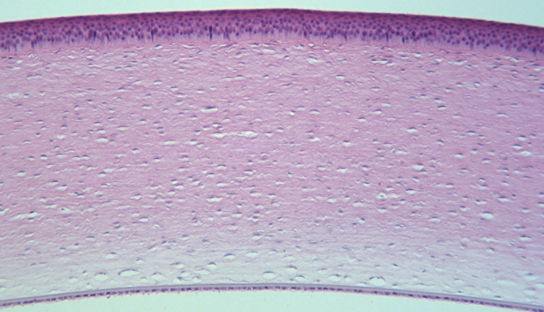
The essential 5 layers of the cornea:
Pre-corneal tear film, corneal epithelium, stroma, Descemet's membrane, and the corneal endothelium.
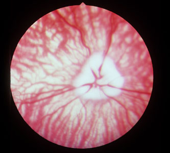
There is a wide margin of variation when it comes to the appearance of the "normal" canine retina.


